

Cone Beam Imaging - Miami, FL
See the details needed for confident dental care
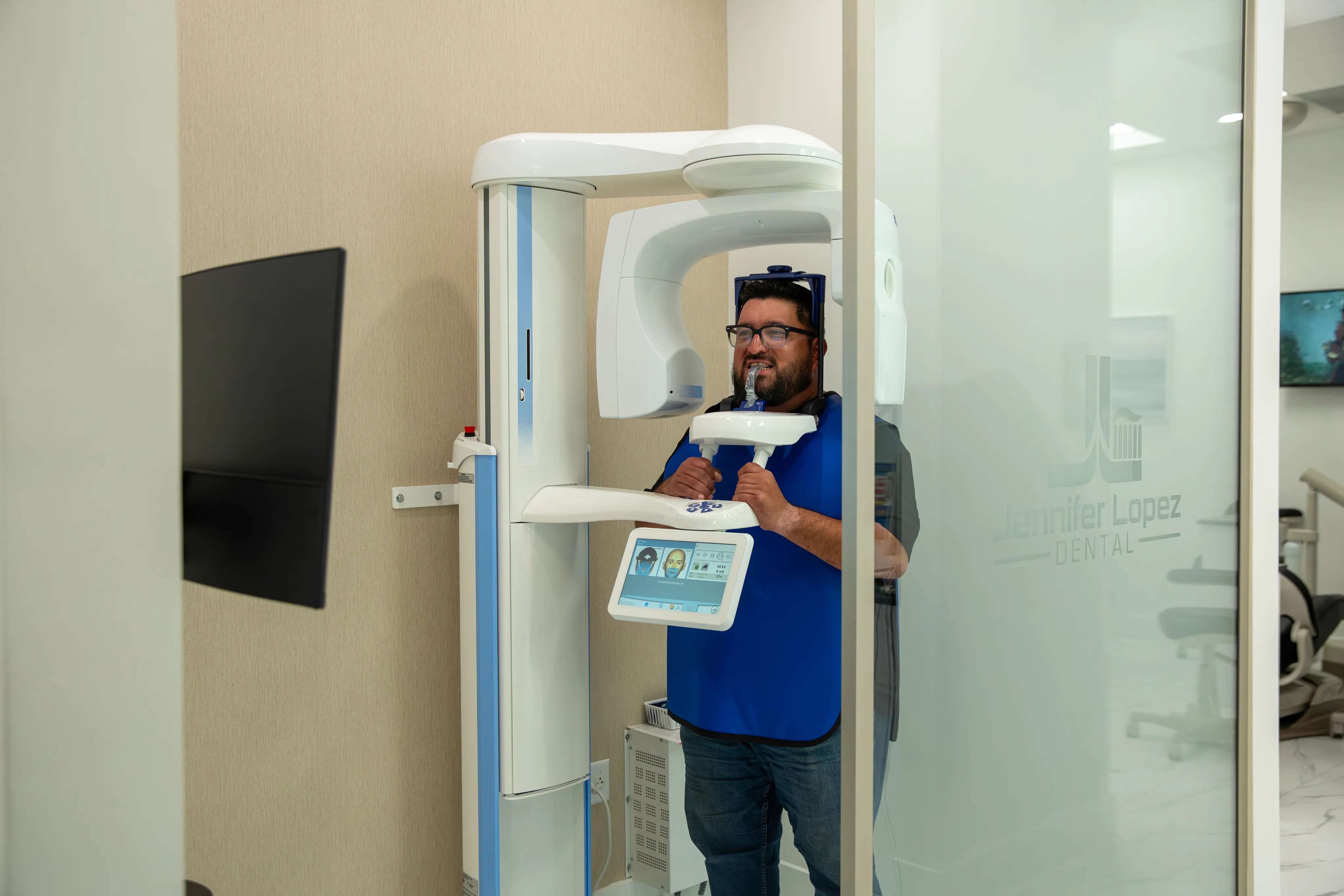
About Cone Beam Imaging
Cone beam imaging is an advanced form of dental X-ray technology. During the scan, the machine rotates around your head and captures hundreds of individual images. Specialized software combines those images into a detailed three-dimensional model that can be viewed from different angles.
The resulting images allow Dr. Lopez to examine teeth, tooth roots, jawbone, nerves, sinuses and other important structures. She can also review individual cross-sections of the scanned area instead of relying only on a flat image.
CBCT differs from a medical CT scan because it uses a cone-shaped X-ray beam focused on the head and neck. Dental CBCT scans also generally require less time and expose patients to less radiation than a conventional medical CT scan.
Benefits of Cone Beam Imaging

Detailed Three-Dimensional Views
CBCT creates a three-dimensional view that can be rotated, enlarged and examined in individual sections.

More Informed Treatment Planning
Detailed images help Dr. Lopez evaluate the position of teeth, roots, nerves, sinuses and bone before recommending treatment.

Precise Measurements
CBCT software allows the dentist to measure bone dimensions and distances between anatomical structures for implant evaluation and more.
Quick, Comfortable Scan
The scanner completes its rotation in approximately 10 to 20 seconds. Nothing is placed inside your mouth during the scan.
Clear Patient Education
Dr. Lopez can review relevant views with you on a screen, making it easier to understand the diagnosis and treatment recommendation.
What to Expect
Your step-by-step guide to cone beam imaging at Jennifer Lopez Dental.
Preparation
You will be asked to remove glasses, earrings, removable dental appliances and other metal objects that could interfere with the images.
Positioning
You will stand or sit inside the scanner. Gentle supports help position your head correctly and keep it still during image capture.
Quick Scan
The scanner rotates around your head once while you remain still. Image capture usually takes about 10 to 20 seconds.
Image Processing
The computer combines the captured images into a three-dimensional digital model that Dr. Lopez can view from different angles.
Review and Discussion
Dr. Lopez reviews the clinically relevant findings and explains how they affect your diagnosis or treatment options.
Frequently Asked Questions About Cone Beam Imaging
How is CBCT different from traditional dental X-rays?
Traditional dental X-rays produce two-dimensional images. CBCT captures a three-dimensional volume that can be viewed from multiple angles and in individual cross-sections. It may reveal details about roots, bone, nerves and sinuses that are difficult to evaluate on a flat image.
Is cone beam imaging safe?
CBCT uses a focused X-ray beam and a short scan time. Radiation exposure is generally lower than a medical CT scan but higher than standard dental X-rays. Dr. Lopez recommends CBCT only when the additional diagnostic information is expected to benefit your care.
How long does a CBCT scan take?
The scanner generally takes about 10 to 20 seconds to rotate around your head. Positioning and preparation require a few additional minutes, but the overall process is quick.
Does a cone beam scan hurt?
No. The scan is noninvasive and does not touch your face. You will need to remain still while the machine rotates around your head.
Do I need to prepare for a CBCT scan?
Most patients do not need special preparation. You may be asked to remove glasses, jewelry, removable dental appliances and other metal objects near the head and neck. You can generally eat, drink and take medications as usual unless instructed otherwise.
When might my dentist recommend CBCT?
Common reasons include implant planning, evaluating impacted teeth, assessing jawbone and sinus anatomy, analyzing root canal anatomy, diagnosing suspected fractures and reviewing TMJ or airway concerns.
Will I need a CBCT scan at every dental visit?
No. CBCT is used when three-dimensional information is needed for a specific diagnostic or treatment-planning purpose. Routine examinations generally rely on clinical findings and conventional dental X-rays.
Can I see my CBCT images?
Yes. Dr. Lopez can display relevant views on a screen and explain the findings. This can help you better understand the condition being evaluated and the available treatment options.

Interested in Cone Beam Imaging?
Let Us Help You
Take the first step toward a healthier, more confident smile. Dr. Lopez and our Miami team will walk you through your cone beam imaging options, answer your questions, and help you make the best decision for your oral health. Call (305) 227-1213 or request an appointment online.
Request a Consultation
Request an Appointment
Fill out the form and we will be in touch shortly.